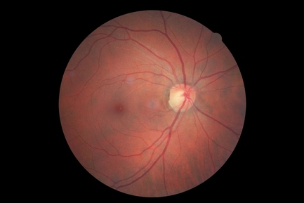
Using a specialized device we take a photograph of the fundus of your eye (retinal image) and we analyze it in depth to detect anomalies. This allows us to give you better explanations and to illustrate our remarks by showing you the photo. We keep the images for comparison during subsequent examinations. This way we observe the evolution of your eyes.